By resurrecting its extinct ancestors, University of Nebraska–Lincoln biologists have helped show that a mutational two-step transformed an overly possessive biomolecule into the four-part, bloodstream-cruising transport that both embraces oxygen and knows how to let it go.
Much is known about vertebrate hemoglobin, the protein in red blood cells that picks up oxygen from lungs or gills and unloads it at tissues throughout the body. Despite the fact that vertebrate animals relied on hemoglobin to evolve their oxygen-fueled metabolism and vastly outgrow invertebrates, though, the evolutionary origins of the protein’s biochemical properties had never been traced.
With the aid of theory, computational models and genetic-engineering tools, researchers from the University of Chicago, University of Nebraska–Lincoln and elsewhere recently managed to do just that. When they did, they discovered that a surprisingly small number of mutations were responsible for the assembly and fine-tuned performance that have made hemoglobin such a critical protein.
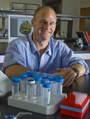
“Hemoglobin provides this nice, simplified system for understanding the evolution of complexity,” said Nebraska’s Jay Storz, Willa Cather Professor of biological sciences. “We were able to distinguish between two competing visions for how that complexity evolves.”
All proteins consist of multi-fold chains that are themselves made from biochemical links, or amino acids. The sequence and interaction of those links determines the structure and behavior of the protein — in the case of hemoglobin, how readily it binds with and releases oxygen. Mutations that swap out certain amino acids for others, then, can sometimes modify the behavior of the entire protein.
Chicago’s Arvind Pillai realized that hemoglobin’s large, varied family — including myoglobin, a single-subunit protein that likewise binds to oxygen but instead stores it in muscle tissue — made it a prime candidate for studying just how incremental or sweeping biomolecular mutations could be. So the team began comparing amino acid sequences from diverse members of the hemoglobin family, then engaged sophisticated statistical methods to infer the sequences of proto-globin proteins in long-extinct ancestors. Among them? The last common ancestor of hemoglobin and myoglobin.
“One way to understand ancestral sequence reconstruction is to think about the evolution of languages,” Storz said. “If we compare the same word in Spanish, Portuguese, French and Italian, since all those Romance languages have Latin roots, we can make some inferences about the specific sequence of letters in the ancestral version of that word.”
Chicago’s Joseph Thornton knew that Storz’s lab had experience synthesizing hemoglobin in cell cultures, purifying those proteins and measuring their biologically relevant properties. As Thornton’s team made progress reconstructing and resurrecting the relevant ancestral proteins, it passed along those proteins to Storz’s, which set out to test the functional properties.
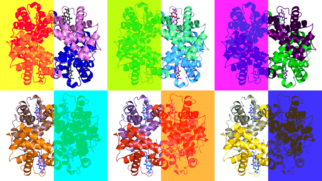
The common ancestor of hemoglobin and myoglobin consisted of just one amino acid chain, the researchers concluded. Eventually, the gene responsible for that ancestral protein was duplicated. That duplicated gene ultimately yielded the one-chain myoglobin before itself diverging, leading to another distinct protein — what the researchers call the “missing link” between the ancient ancestor and hemoglobin.
When Storz and Nebraska postdoctoral researcher Anthony Signore tested the resurrected protein, they confirmed that changes in its surface architecture allowed it to bind with an identical subunit chain, forming a two-subunit complex. But testing the resurrected complex also demonstrated that it clutched oxygen too tightly to ever let it go, making it incapable of delivering that oxygen to cells. And even when given ample opportunity to pair up with other two-subunit complexes and form a four-subunit architecture similar to that of modern hemoglobin, it never did so.
Fortunately for future vertebrates, the essential catch-and-release capacity was on its way. Modern hemoglobin consists of four total chains — two alpha, two beta — that bind together and boast a Goldilocks grip: not too loose, not too tight. Those chains also exhibit a follow-the-leader form of cooperation, with the other three tending to pick up or dispense oxygen whenever one begins doing so.
The researchers had reasoned out that the two-part predecessor of hemoglobin genetically branched into versions of the alpha and beta chains. But they also knew that some further mutations, in one or both chains, must have promoted their aggregation into modern four-chain hemoglobin. As many as a few dozen mutations could have been collectively responsible.
Yet after narrowing down suspects and modeling the behavior of the subunits with various mutations, the team concluded that just two — both along the surface of the beta chain — encouraged the beta to bind with the alpha and, in turn, the alpha-beta duo to bind with another. The team also discovered that the four-subunit assembly just so happened to calibrate hemoglobin’s ideal oxygen grip and promote its four-quadrant cooperation via long-range interactions among amino acids in remote parts of the protein.
“In principle, that kind of increasing complexity could involve many, many, many changes at many different sites in the protein, where every individual change confers just a slight increase in the ability for the subunits to associate,” Storz said. “At the other end of the spectrum, it’s also conceivable that the same property could have evolved in a pretty simple manner, where just one or a few changes were sufficient to confer the ability of the subunits to self-associate and function as part of a multi-subunit assembly.
“What we found is more consistent with the latter scenario, where, surprisingly, a pretty small number of mutational changes were necessary and sufficient to confer this ability.”
Still, Storz emphasized that the series of mutations occurring earlier in hemoglobin’s evolutionary history laid the roots for new functional properties to emerge.
“Basically, the existing structure of the protein at a given point in time dictated what changes were possible and which changes could potentially confer a new property,” he said. “If that structure had been different, then other changes would have been required to confer that same property.
“It looks like the particular paths that evolution follows are always very strongly influenced by the ancestral starting point.”
And if that evolutionary serendipity hadn’t played out the way it did? If prior mutations hadn’t aligned with and toppled those final two dominoes?
“Without some chemical means of transporting oxygen in a closed circulatory system,” Storz said, “vertebrate evolution would have looked very, very different.”
The team reported its findings in the journal Nature. Thornton, Pillai, Storz and Signore authored the study with UChicago’s Georg Hochberg and Carlos Cortez-Romero; the University of Oxford’s Shane Chandler and Justin Benesch; and Texas A&M University’s Yang Liu and Arthur Laganowsky. The researchers received support from the National Institutes of Health and the National Science Foundation.