· 3 min read
Chemists measure aggregation of disease-related proteins
Predators looking to snack on deep-sea shrimp sometimes encounter a surprise: clouds of blinding blue light that their prey spew as a distraction before bolting for new hiding spots in the jet-black depths.
A new study from the University of Nebraska-Lincoln has shown how the enzyme that sparks these bioluminescent fireworks can be used to monitor an indicator of human diseases that include Alzheimer’s, Parkinson’s and Type 2 diabetes.
The new screening technique uses self-assembling fragments of the enzyme to measure a potentially toxic process known as protein aggregation in living cells. Using its new system, the UNL research team hopes to see the light: The less aggregation occurring within a cell, the brighter the system glows blue.
As workhorses of the cell, proteins naturally fold into specific three-dimensional forms that dictate their functions. However, these proteins sometimes misfold – the biological equivalent of an origami crane mutating into a bulldog – which leads them to clump together, or aggregate. Previous research has linked the protein aggregation process with multiple diseases.
Assistant professor Cliff Stains and his colleagues demonstrated that their technique can quickly test compounds that might prevent this aggregation and potentially combat the diseases associated with it. The technique could potentially screen tens of thousands of compounds in a single day, Stains said.
“The idea is that we’re going to generate genetically encodable libraries of a few hundred thousand compounds,” said Stains. “Then we’re going to screen these compounds against aggregating proteins to see if we can find any that decrease the aggregation potential.
“We can visualize tens of thousands of cells on a plate and just look for the most luminescent one. This will allow us to screen really large compound libraries pretty rapidly.”
Unlike many of its predecessors, the team’s approach can accomplish this within living mammalian cells – an important advancement that offers a better sense of how drug-like compounds might behave in a biological setting, Stains said.
The team’s technique involves splitting a bioluminescent enzyme into two pieces, significantly reducing its luminescence. Given the opportunity, however, these enzyme fragments will spontaneously snap back together and shine just as brightly as before.
Knowing that protein aggregation interrupts this reassembly, the researchers attached one enzyme half to an aggregation-prone protein. When that aggregation occurs, the lights remain dimmed; when it doesn’t, the blue luminescence returns.
The result: A hyper-efficient, intuitive readout system that allows the researchers to identify molecules that reduce the aggregation potential of a protein.
The system also serves as a convenient platform for addressing important questions about the protein aggregation process, according to Stains. The team has already begun triggering random mutations in proteins of interest and identifying those that lead to aggregation.
“What residues in a protein are really essential for aggregation? What is the mechanism (that drives) aggregation? We can ask those kinds of fundamental science questions with our new technique,” Stains said.
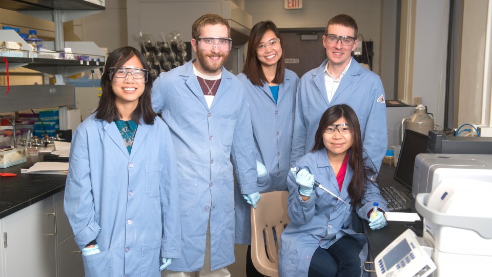
The team’s study appeared in the journal ACS Chemical Biology. Stains co-authored the paper with doctoral students Jia “Emma” Zhao and Travis Nelson, who is a fellow of UNL’s Molecular Mechanisms of Disease program. Undergraduate students Quyen Vu and Tiffany Truong also contributed to the research through UNL’s UCARE Program, with Truong receiving the support of a Distinguished Life Sciences Scholarship.

Recent News